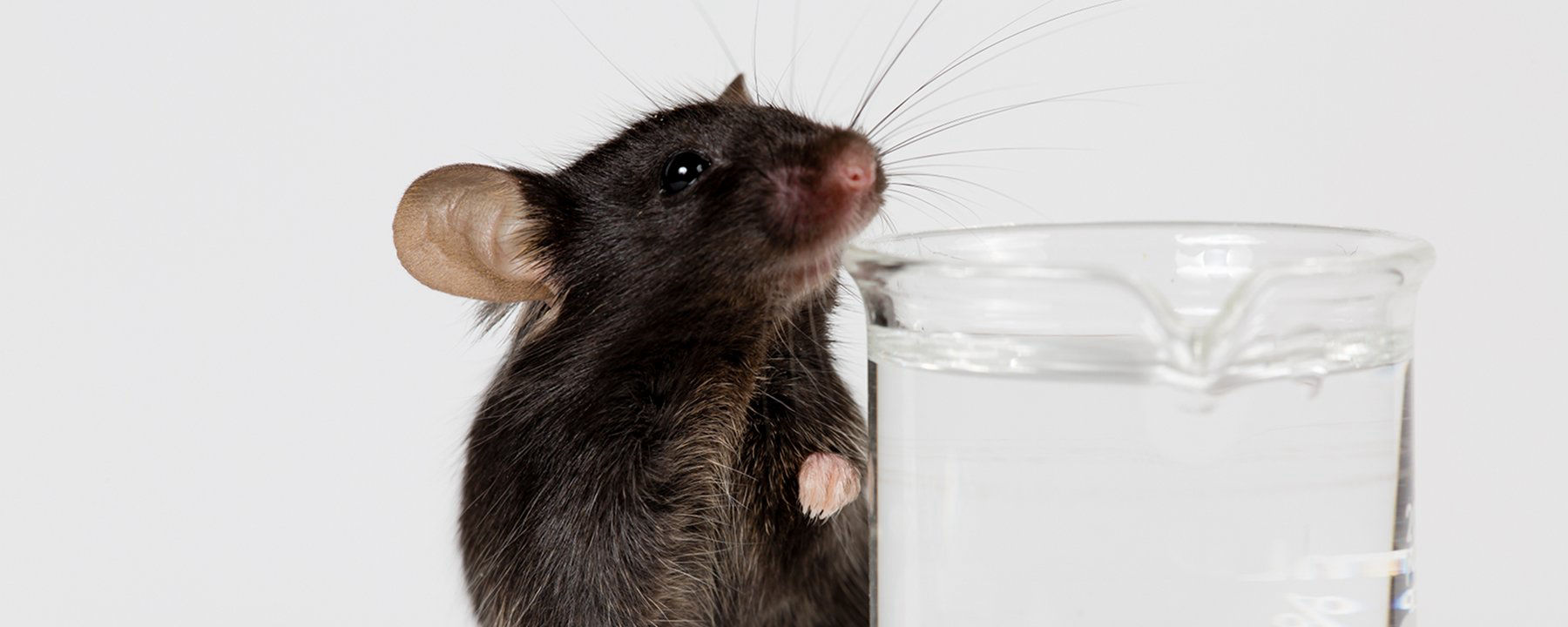
Electrical recordings of approximately 24,000 individual neurons across 34 regions of the mouse brain reveal, in a study published in Science today (April 4), the cells that become activated during thirst, drinking, and satiety. The results show the widespread distribution of neuronal activity at different phases of the process and how these patterns of activity can be largely recapitulated by the stimulation of a specific group of sensory cells.
“[The work provides] a very detailed look at one of the most basic processes that terrestrial animals need to be able to do in order to stay alive,” says neurobiologist Scott Sternson of the Howard Hughes Medical Institute’s (HHMI) Janelia Research Campus who was not involved in the research.
“It’s really a tour de force that they were able to record from so many neurons,” adds neurologist Charles Bourque of McGill University who also did not participate in the work.
Physiological signals related to dehydration, such as sodium levels and blood osmolarity, are detected by a small group of sensory cells in a region of the brain called the subfornical organ (SFO). These cells are critical for the sensation of thirst and the subsequent motivation to drink, and have even been shown, when artificially activated, to induce thirst-like behavior in fully hydrated animals, says physiologist and HHMI investigator Zachary Knight of the University of California, San Francisco.
Just how the natural or artificial stimulation of SFO neurons leads to the subsequent activation and coordination of downstream neural circuitry to produce motivation and behavior—thirst and drinking—is largely unknown.
To investigate these downstream events, Karl Deisseroth of Stanford University and colleagues examined brain-wide neuronal activity in thirsty mice using Neuropixels probes. These newly developed electrophysiological devices consist of nearly 1,000 recording sites along a thin shank less than a tenth of a millimeter thick that can be inserted into the brain of a mouse with minimal damage, allowing for simultaneous recordings of hundreds of single neurons at a range of depths. These probes enabled the team to record 23,881 neurons during 87 separate sessions that probed 34 different brain regions in 21 mice.
The recordings were performed in thirsty mice whose heads were fixed in position and that had been trained to respond to two different odor cues—one which meant water was available in a spout if they licked it, the other that signaled water was not available. The recording sessions covered the entire process from thirst through drinking on cue to satiety. Despite the deep insertion of the probes in their brains, “animals with these electrodes [in place] are healthy, non-distressed, and learn as rapidly as if there were no probe,” writes Deisseroth in an email to The Scientist.
The team’s analysis of the resulting data revealed that a large proportion of the neurons in all brain regions probed were activated both in response to the cue and in the subsequent task of drinking. It was “a big surprise,” writes Deisseroth, that “even for a task as simple as a thirsty mammal seeking water, most of the brain, and most of the corresponding neuronal population, becomes involved in the task.”
The data also revealed that patterns of cell activity mainly fell into three groups: those whose activity depended on the underlying physiological state of the animal (either thirsty or sated); those whose activity depended on the particular cue given; and those whose activity depended on behavior (licking or not). While, largely speaking, each neuron fell into one of these categories, each brain region contained a mix of the three.
The team went on to show that optogenetic stimulation of the SFO neurons in fully sated animals could not only restore behavior indicative of thirst (as previously shown), but also spark the neuronal activity patterns exhibited by the animals when they had been thirsty.
“That’s the exciting thing,” says Knight, “that you can take a small population of sensory neurons in the SFO—just a few thousand cells—stimulate them, and change global brain dynamics. . . . [The study] just underscores how powerful these cells are.”
While the paper largely frames the results in terms of these general observations about types of neuronal activity, it also provides a wealth of more-specific data in the form of the thousands of individual recordings from particular brain sites.
“There had been relatively little known about the activity at the individual-neuron level across so many different brain regions,” says Sternson. “What this study has created is a lot of new knowledge,” and that “will be very helpful to the field going forward.”


RSS